Multiphoton Microscopy
Democratizing deep 3D live imaging
Power of deep-tissue multiphoton imaging,
at your benchtop with accessible pricing
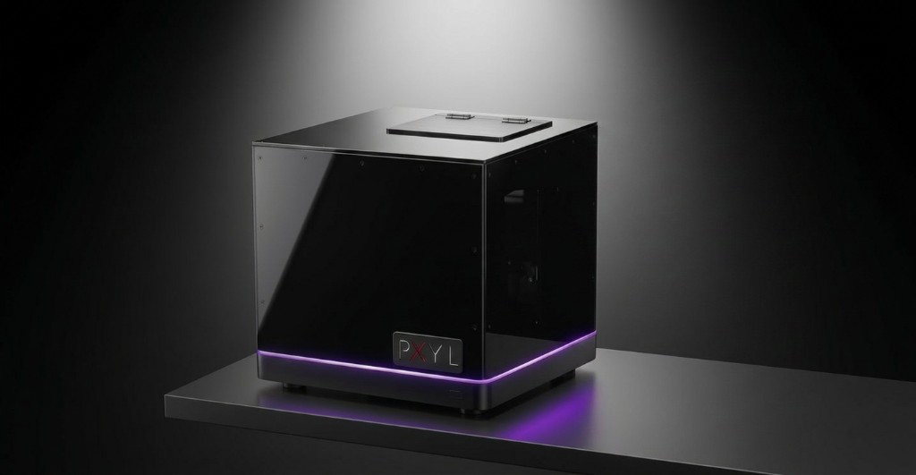
Technical Capabilities
PXYL combines multi-modal optical imaging and validated AI inference in a compact bench-top form factor — no optical table or darkroom required
Operates effortlessly on a standard lab bench. Reclaim your lab space with a system that requires no specialized infrastructure
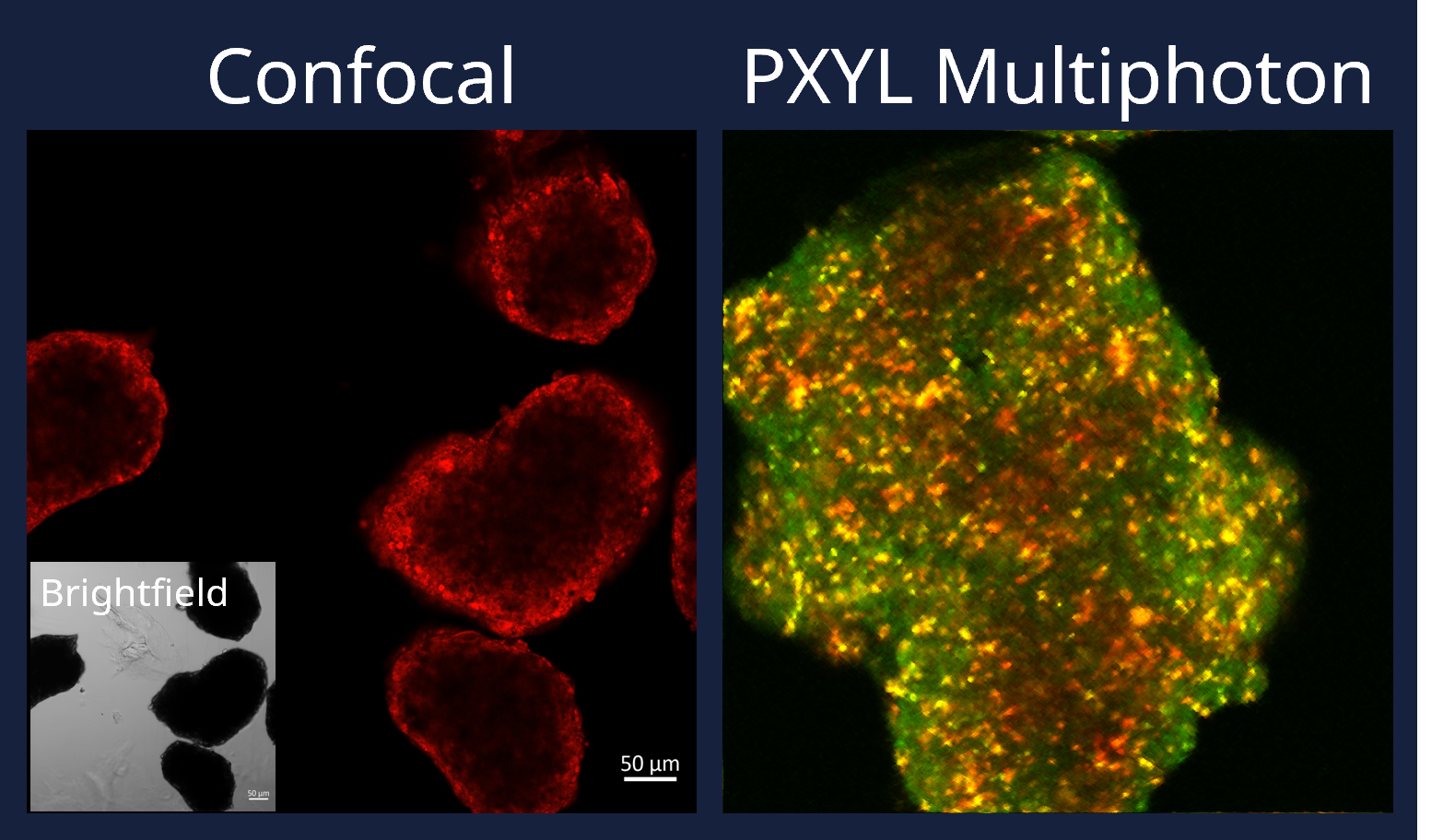
Achieve up to 500 µm penetration depth with incredibly low phototoxicity. Optimized for long-term, true-to-life live cell observation
Custom-engineered with a high NA (>0.65) and an expansive 4 mm working distance. Seamlessly compatible with multi-well plates and microfluidics
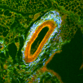
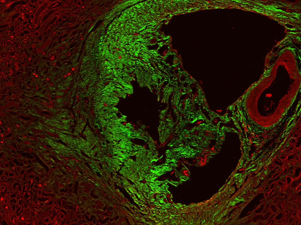
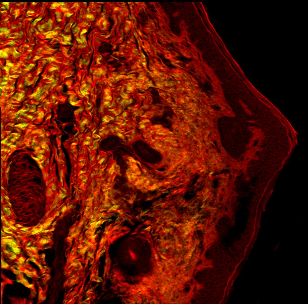
Unlock comprehensive tissue insights. Capture 2PEF, SHG, THG, and label-free metabolic imaging (NADH/FAD)—all within a single, unified architecture
A true turnkey solution. PXYL’s intuitive software streamlines your entire workflow, from setup to complex data acquisition
Advance your biological studies. Move fluidly from high-resolution 3D spatial mapping to complex 4D time-lapse imaging
System Comparison
| Feature | PXYL MPM | Commercial MPM | Typical Confocal |
|---|---|---|---|
| Primary Application | Deep, Live 3D/4D Imaging | Deep, Live 3D/4D Imaging | High-Resolution Surface Imaging |
| Imaging Depth | Up to 500 µm | Up to 500 µm | ~100–150 µm |
| Phototoxicity | Very Low | Very Low | High |
| Working Distance | Up to 4 mm | Typically <1 mm | Typically <1 mm |
| Sample Versatility | Slides, Well-Plates, Microfluidics | Slides, Dishes (limited by WD) | Slides, Dishes (limited by WD) |
| System Footprint | Compact Benchtop | Optical Table & Dark Room | Optical Table & Dark Room |
| Acquisition Cost (USD) | Most affordable available | Very High | High |
Potential Applications
Track growth deep inside 3D organoids over days, not minutes. PXYL's gentle, deep penetration (up to 500µm) and extra-long working distance enable continuous low-toxicity imaging for organ-on-chip systems, while visualising the microenvironment and collagen scaffolds (SHG) without destroying your sample.
"See the structure. Understand the function. Keep it alive."
A versatile bioimaging tool for diverse research applications. Capture multiple contrast modes—from immunostained cells in thick tissue to label-free NADH/FAD metabolic imaging and SHG/THG structural contrast. Image up to 6 channels simultaneously for comprehensive multi-modal analysis in a single, compact system.
"Pixel-perfect multi-channel contrast, deep within the tissue."
PXYL eliminates the traditional barriers to multiphoton microscopy, making deep tissue imaging accessible to every research lab.
One of the most affordable multiphoton platforms available
Delivering a 5–10× cost reduction compared to traditional systems
Fits on any lab bench — no optical table or darkroom required
4mm working distance enables imaging in multi-well plates and organ-on-chip devices
Up to 500µm imaging depth with minimal phototoxicity for long-term live studies
PXYL eliminates the traditional trade-offs. Uncompromised, commercial-grade multiphoton performance—finally accessible for your bench and your budget