Medical Device

Glanzir®

Ushering pathology into the "Slide-Free" Era
From fresh tissue to digital H&E: in just 3 minutes

Enquire Now
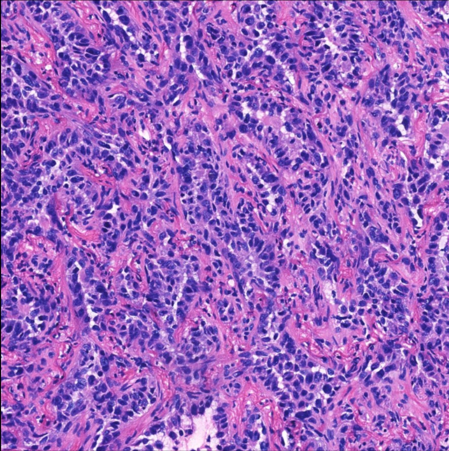
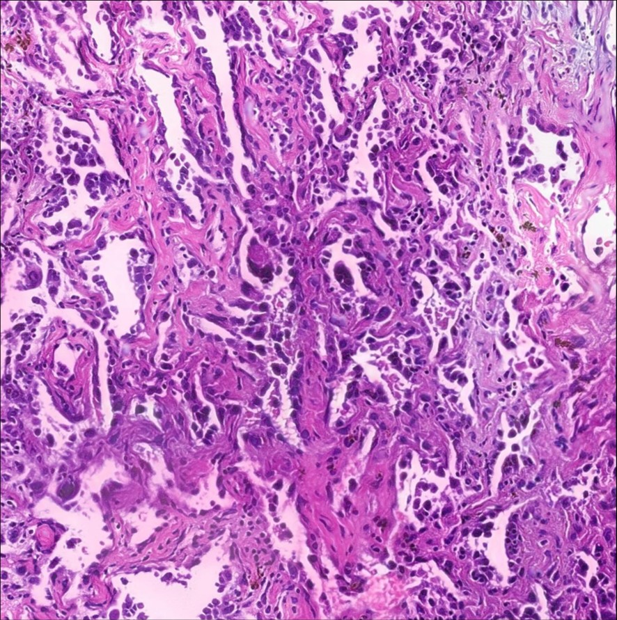
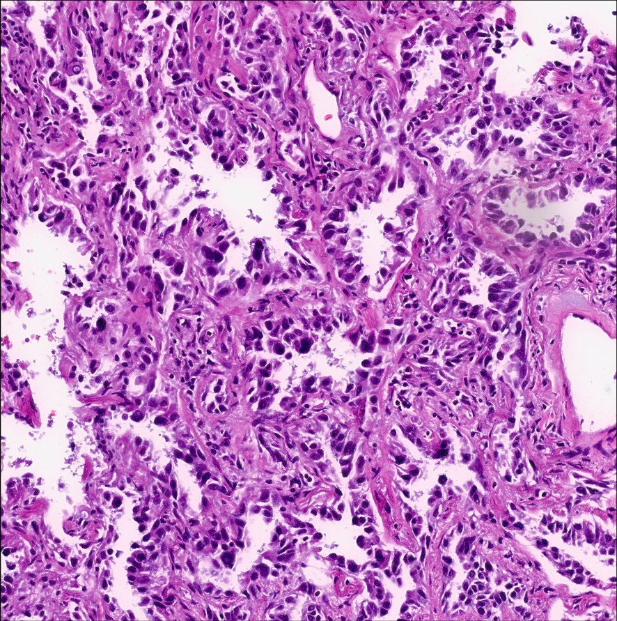
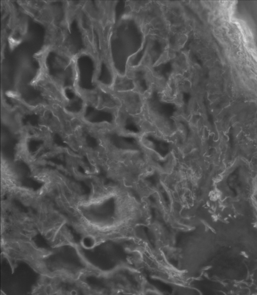
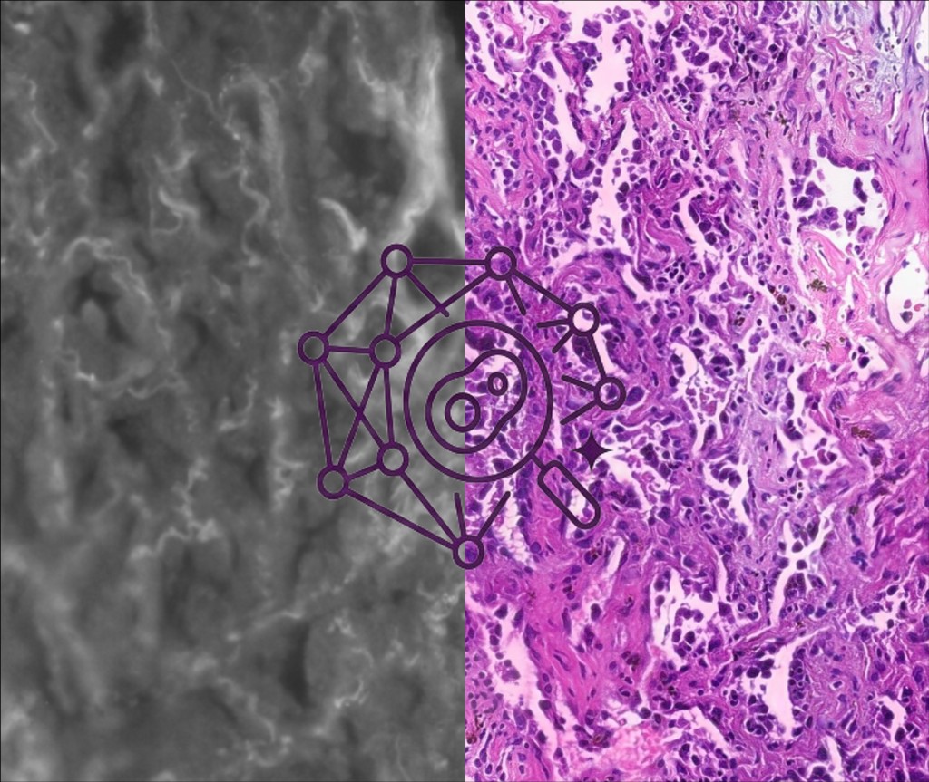
Slide-Free & Stain-Free Unlocking the era of digital pathology for fresh tissue
3-Minute Rapid Imaging Fulfilling intraoperative requirements effortlessly and reducing labor-intensive tasks
Computational H&E Imaging High-fidelity reconstruction of morphological and histological details

Workflow Comparison

From Anxiety to Efficiency

The Past

The conventional intraoperative workflow is labor-intensive, requiring multiple manual steps including embedding, sectioning, fixation, and staining. Because slide quality is highly operator-dependent and diagnostic accuracy relies heavily on individual experience, pathology departments face immense pressure, leading to prolonged intraoperative wait times for surgical teams.

The Future

Glanzir® bridges the gap from fresh tissue to digital "Computational H&E" images in just 3 minutes. By providing a rapid, high-fidelity, and non-destructive diagnostic paradigm, Glanzir ensures clinical precision while significantly accelerating surgical turnover and liberating pathology staff from manual bottlenecks.

Clinical Trial

Image Comparison

Interoperative Frozen Section

Frozen sectioning is the current clinical routine for intraoperative pathology. It typically requires 30 minutes and often suffers from significant image degradation compared to the FFPE gold standard.

Frozen section sample

abdc

Glanzir® Scan

Enabling direct digitization of fresh tissue into computational H&E images. The image quality surpasses that of traditional frozen sections, offering a closer approximation to the FFPE gold standard.

Glanzir scan sample

abdc

FFPE (Gold Standard)

H&E staining following paraffin sectioning is the gold standard for pathological examination, and normally takes more than 3 days.

H&E stained sample

abdc

Core Technology

Technical Excellence:
Autofluorescence Imaging (UV-AF)

Glanzir revolutionizes traditional tissue processing by capturing "native signals" directly from the tissue surface through two core technological leaps:

Endogenous Autofluorescence Imaging

Slide-Free Imaging: Excitation light penetrates only the tissue surface to capture 2D images, eliminating signal interference from deep tissue.
Stain-Free: By utilizing Deep-UV light to excite endogenous fluorophores, high-contrast imaging between the nucleus and cytoplasm is achieved without any chemical labeling.

UV fluorescence microscopy
H&E color reconstruction

Computational H&E algorithm

By leveraging endogenous autofluorescence and tissue morphology, our system precisely maps signals into the traditional H&E color palette. This enables pathologists to interpret images intuitively, removing diagnostic barriers.

Objective 20×
Resolution 0.55 μm/pixel
Dimensions (L×W×H) 600×600×320 mm
Max Sample Size 25×25×10 mm

Strategic Roadmap

Future-Proofing Pathology

Expanding clinical coverage across critical cancer types

Now

Currently Supported

Glanzir is clinically validated and actively deployed for intraoperative assessment of lung and breast cancer cases.

Lung Breast
Upcoming

Expanding Coverage

Active clinical validation and algorithm development underway to extend Glanzir's diagnostic capabilities across five additional cancer types.

Thyroid Prostate Ovary Cervix Colorectal
Future

Broader Horizons

Further oncology indications and new clinical applications under research. Contact us to discuss partnership opportunities.